Tumor cells divide rapidly. That is why chemotherapy is often the first-line treatment for many types of cancer. Chemotherapy specifically targets rapidly dividing cells, with the goal of stopping tumor growth by killing tumor cells.
The problem is that some tumor cells are not susceptible to chemotherapy. This occurs, for example, in ovarian cancer, where more than half of patients are resistant to chemotherapy. These patients only experience side effects, while the tumor continues to grow. Why does this happen? Molecular geneticist Nitika Taneja is searching for the answer.
One explanation for the immunity of some tumor cells likely lies in the way the DNA divides in those cells. Some of these cells pack their DNA very tightly during cell division. As a result, they are less sensitive to damage caused by chemotherapy. This tightly coiled DNA is called heterochromatin. The amount of heterochromatin is an indicator of how susceptible a cell is to chemotherapy.
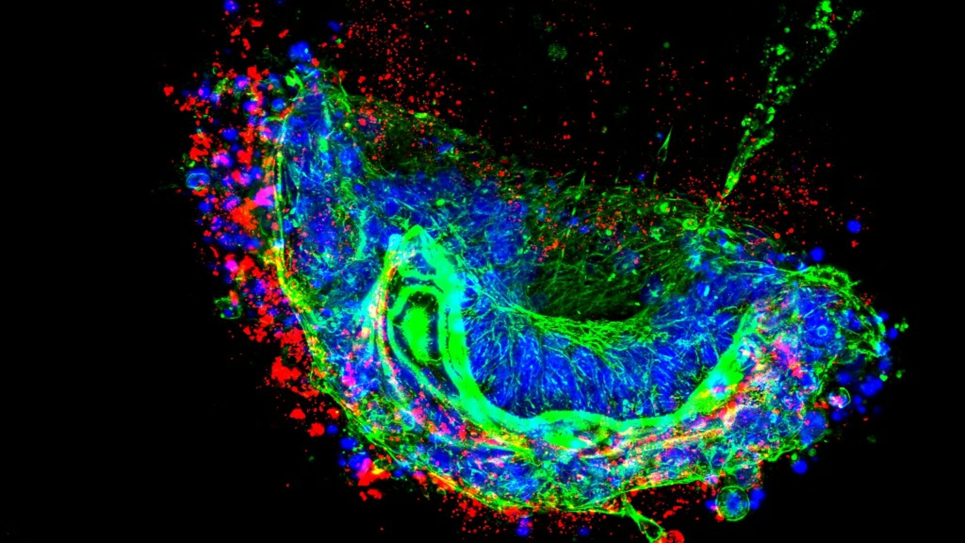
DNA on a microscope slide
Nitika and her colleagues used this knowledge to develop their own technique that allows them to accurately determine the amount of heterochromatin in DNA during replication. Achieving precise visualization is not straightforward due to the properties of the tissue.
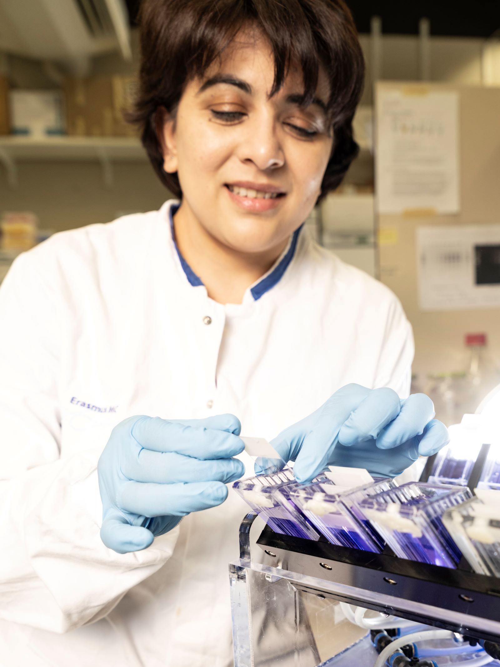
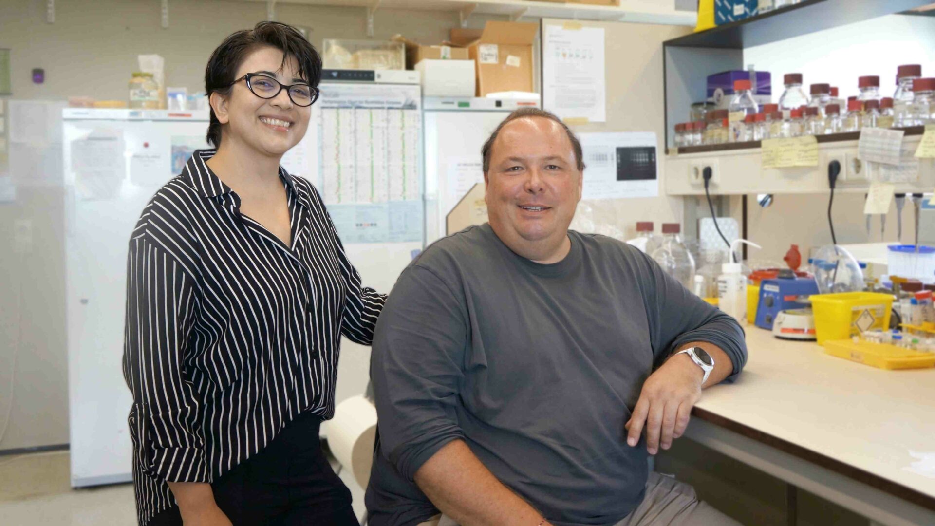
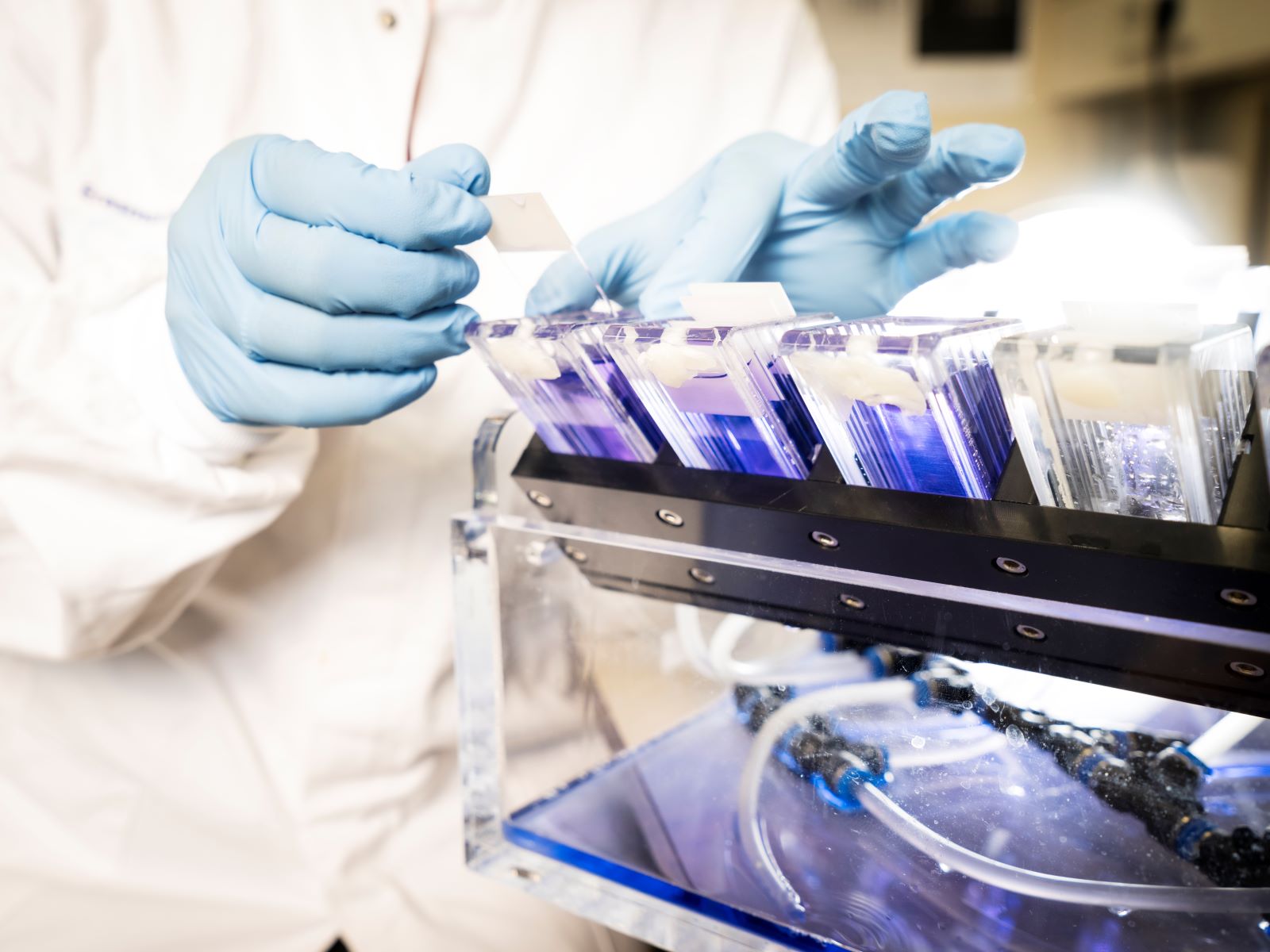
Nitika and the ChromStretch, photo by Harmen de Jong
DNA and its associated proteins tend to clump together rapidly. ‘In such a tangled mass, it is impossible to determine the composition and thickness of individual fibers.’ To overcome this, they built a device that can spread DNA from a cell onto a microscope slide very precisely, without clumping.
The prototype of their technique, called ChromStretch, was developed by the Department of Experimental Medical Instrumentation at Erasmus MC. ‘The first version consisted of test tubes, tubing, and an aquarium pump we bought online for twenty euros.’
Despite this improvised setup, the method worked well. ‘The liquid containing the DNA does not drip, but flows very gradually. The device operates at exactly the right angle, force, and speed. As a result, the fibers remain separated, allowing us to examine them properly under the microscope.’
Testing on cancer cells
Before ChromStretch can be used routinely, further work is required. Nevertheless, Nitika and her colleagues are already making significant progress. In fact, they have established a collaboration with the Department of Medical Oncology to test the technique on cancer cells.
‘The first version consisted of test tubes, tubing, and an aquarium pump we bought online for twenty euros.’
‘We receive tissue samples from patients with ovarian cancer, half of whom are known not to respond to a specific type of chemotherapy.’ By treating these cells with chemotherapy and then analyzing the DNA, Nitika can observe what goes wrong during the replication process. ‘Within a few days, we can determine whether a patient is susceptible to this specific type of chemotherapy. This way, we hope to predict how the patient will respond to the therapy.’
The research is still ongoing. Although the results are promising, the current study involves only 25 patients. Thanks to a grant that Nitika received with John Martens and Ingrid Boere, the study can be expanded to 100 patients.
Unzipping DNA
What exactly is heterochromatin, and why might it play an important role in cancer treatment? To understand this, we return to the basics: a dividing cell. During cell division, DNA is duplicated and split into two. The double helix must first partially unzip. The unzipped section of DNA is called the replication bubble. The junction where the DNA unzips, takes on a Y-shaped form. ‘That structure is known as the replication fork’, says Nitika. ‘This is where DNA is most vulnerable.’
It is at this stage of cell division, at the replication fork, that chemotherapy is most effective. The treatment works by damaging the DNA strand or halting the replication process. The more damage a cell accumulates, the harder it becomes to repair. If repair fails, the cell dies. In theory, this is the ideal way to treat cancer.
In practice, however, chemotherapy does not affect all cancer cells. Some of these cells package their DNA at the replication fork in a slightly different way. The DNA is wound more tightly, making it less susceptible to damage. ‘Researchers previously believed that DNA is only tightly packed when it does not need to be read. However, we have discovered that this winding is a way for the cell to protect the DNA.’
This compact DNA structure reflects how a cell preserves its genetic information and protects it from external influences. One cell is better at this protective mechanism than another. That is why most healthy cells are relatively effective at protecting their DNA.
Tumor cells usually contain mutations and are less capable of repairing damage, making them more sensitive to chemotherapy. However, some ‘super tumor cells’ behave differently. They contain so many mutations that they have become stronger as a result. These cells can produce large amounts of heterochromatin at DNA replication sites, enhancing their ability to protect DNA.
ChromStrech, photo by Harmen de Jong
Using their technique, Nitika and her colleagues aim to understand how these ‘super tumor cells’ generate so much heterochromatin. What exactly does heterochromatin do, and can its formation be inhibited?
New drugs
They have already discovered that certain proteins are overrepresented in resistant tumor cells. These proteins can form loop-like structures around replication forks, further protecting DNA. Some of these proteins may serve as targets for new drugs. Although several candidate compounds are in early development, much remains unknown about their effects. ‘These drugs could be toxic, because healthy cells also contain these proteins. This requires careful investigation.’
Nitika’s immediate goal is to understand how cancer cell DNA replicates and protects itself. ‘Therefore, we compare sensitive and resistant tumors using ChromStretch. Changes at the level of replication forks, such as heterochromatin formation, can provide insights into the expected effectiveness of chemotherapy. This knowledge can help distinguish between cancer types and improve predictions of treatment response.’
Ultimately, Nitika hopes to contribute to more personalized treatment strategies. Better-matched therapies could reduce unnecessary side effects and improve patient outcomes.